This page is not compatible with Internet Explorer.
For security reasons, we recommend that you use an up-to-date browser, such as Microsoft Edge, Google Chrome, Safari, or Mozilla Firefox.
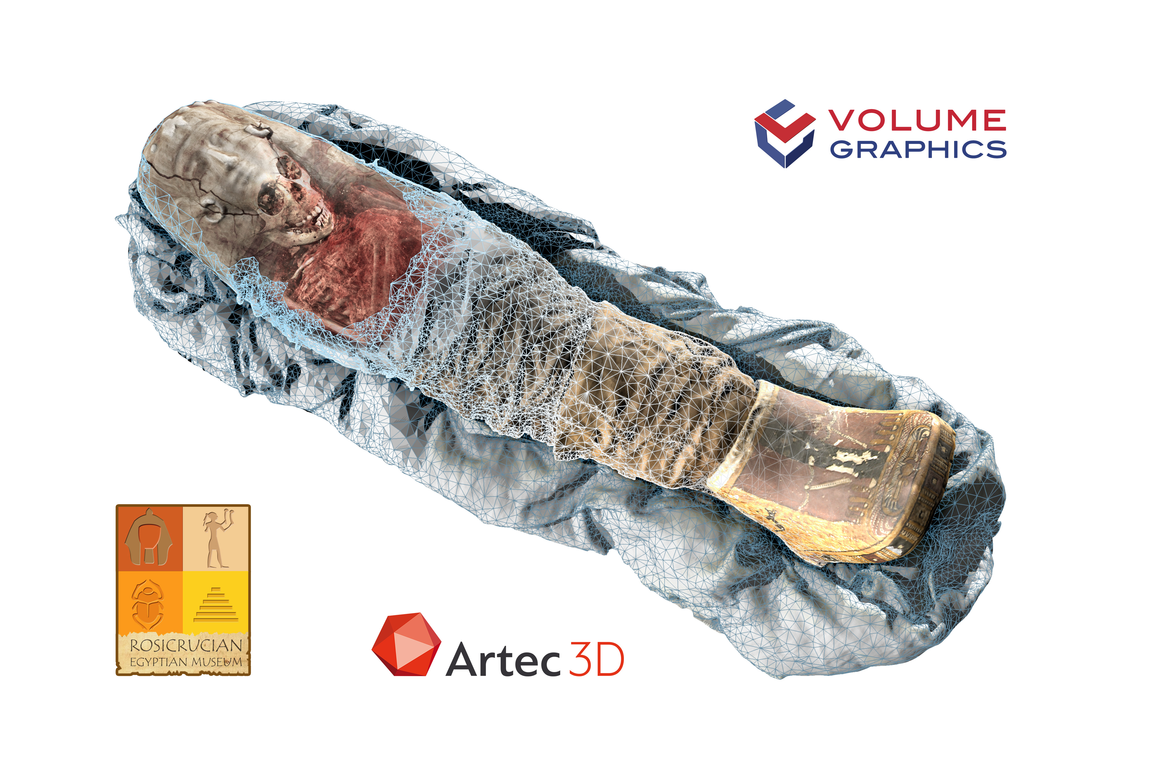
The mummy "Sherit" as a combined representation of CT and optical scans. The light blue mesh is for illustrative purposes only; it symbolizes the now possible combination of colored 3D surface scans and the internal three-dimensional data obtained by a CT scan.
How a Mummy Got Its Color Back
A new software feature from Volume Graphics allows you to combine the visually powerful data of an optical 3D scanner with the all-encompassing detail of a CT scan with just one click. One of the first applications of this new feature was on the scan of the Sherit mummy.
Sherit, ancient Egyptian for "little one," is a mummified Egyptian child who died two millennia ago at the age of 4. It currently resides at the Rosicrucian Egyptian Museum in San Jose, California.

A high precision optical scan, taken with a handheld 3D scanner by Luxembourg company Artec 3D, gave an impressively vivid, full-color 3D image of the surface of the mummy. The 3D image is stored as a textured mesh, which contains both color and geometrical information. In addition, a CT scan created with an AXIOM Siemens scanner detected the complete internal structure of the mummy non-destructively, an area which remained hidden to the optical scanner. A three-dimensional volume was calculated from the CT scan and included all material, tissue, and geometric information.
Then, all that was left to do was combine both scans into a single 3D model. For this, the textured mesh was placed on the surface of the object calculated from the CT scan. The result was, in this case, a mummy that is the most exact digital copy of the original to date, both within and without.
While CT delivers comprehensive material information, no color information is included. Previously, CT-scanned objects had to be manually colored in order to give the surface a realistic look. Thanks to Volume Graphics software, this step is no longer required if you also perform an optical scan of the object in addition to the CT scan. The function to read OBJ and PLY data is part of the new version VGSTUDIO MAX 3.1. Volume Graphics software supports colored point clouds as well as meshes. VGSTUDIO MAX is the high-end software for the visualization and analysis of industrial computed tomography (CT) data.
Christof Reinhart, CEO of Volume Graphics: “We’re very thrilled to see what the users of our software will do now that they’re able to add another dimension to their volume data! By adding colored point clouds as well as meshes, the previously separated worlds of optical and CT scanners are merging. This allows for a more life-like, accurate representation of all kinds of objects and thus improves our understanding of these scanned objects.”
Obvious fields of application are protection of cultural heritage, forensics, archeology, anthropology, as well as medical sciences. However, there are possible industrial applications, as well. For example, the combination of textured meshes with CT can be used to examine details on the inside of an object in context with color details on the surface.